




Sophont has published the Holon-S1 technical report, a model that tries to read gene activity off an ordinary pathology slide. The report on Sophont's site is undated beyond 2026, and the model started circulating widely on October 20. The authors are Hassan Muhammad, Benjamin Warner, Paul S. Scotti and Tanishq Mathew Abraham. Sophont describes Holon-S1 as predicting "spatially resolved genomic profiles, signalling pathways, and biological programs" from H&E morphology, and states plainly that it is "currently a research-only tool and not yet approved for patient diagnosis."
The reason it matters: a biopsy almost always yields an H&E slide, but only a few labs can afford spatial transcriptomics, the assays that show where each gene is switched on inside a tumor. If a model can approximate the second from the first, spatial molecular information becomes cheap. Whether that approximation is good enough is exactly what the report tries to measure.
TL;DR: Holon-S1 at a glance
| Question | Answer |
|---|---|
| What does it take as input? | A routine H&E whole-slide image |
| What does it output? | Spatially resolved gene expression, pathway activity and biological programs |
| Who built it? | Sophont; authors Muhammad, Warner, Scotti, Abraham |
| Is it clinical? | No. Research-only, not approved for diagnosis |
| Reported comparisons | Beat DeepSpot-M on Atera data; higher median correlation than reported Path2Space on a curated subset |
| Tissue types | FFPE (n=8) and fresh-frozen (n=31) slides did not differ significantly (p = 0.184) |
| Biomarker hints | EREG gradient in triple-negative breast cancer; proliferation versus stroma/EMT segregation in colorectal cancer |
| Biggest caveat | Retrospective, regional (not single-cell), mixed assay platforms |
What problem does Holon-S1 solve?
Pathology has two views of the same tumor. H&E staining shows morphology, the phenotype. Spatial transcriptomics (ST) shows genotype in space: which genes are active, and where. The two are different windows onto one system, as Sophont's own figure shows, and ST platforms differ in resolution, noise and panel size.
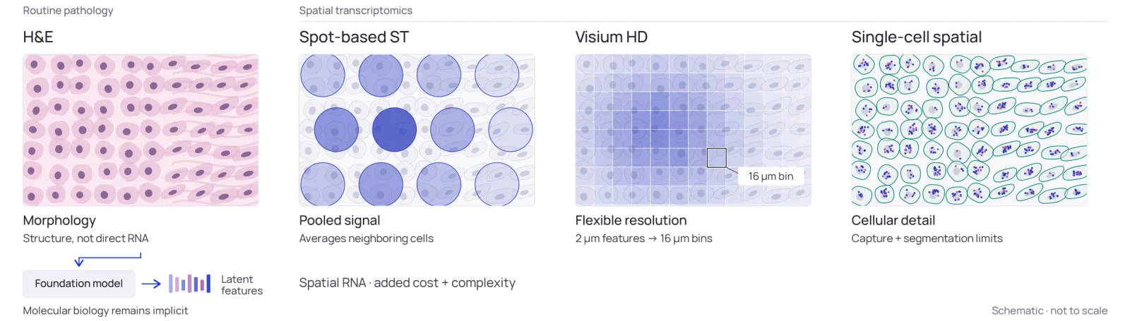 Holon-S1 spatial gene expression prediction: H&E morphology versus spatial transcriptomics platforms
Image: Sophont
Holon-S1 is trained pan-cancer on data from several ST platforms and resolutions, including spot-level and single-cell measurements. It combines fine cellular morphology with broader slide-level spatial context, then predicts expression across the slide. The report does not describe the architecture, parameter count or training-set size in the page we read, so those details are unknown to us.
How good are the predictions?
Sophont reports several comparisons. All of them are the authors' own, and the model has not been independently replicated in anything we found.
- Platform effect. Predictions scored higher against newer platforms. The Atera platform gave a higher median Pearson correlation than any Xenium sample.
- Versus DeepSpot-M. On Atera whole-transcriptome data, Holon-S1 outperformed DeepSpot-M, even though it had significantly fewer breast-tissue samples in training.
- Versus Path2Space. On a quality-curated subset of the HTAN dataset, Holon-S1 reached a higher median correlation than Path2Space's reported numbers (not a head-to-head run), and it performed comparably on slides Path2Space excluded.
- Tissue preparation. FFPE and fresh-frozen slides did not differ significantly (p = 0.184), which matters because clinical archives are mostly FFPE.
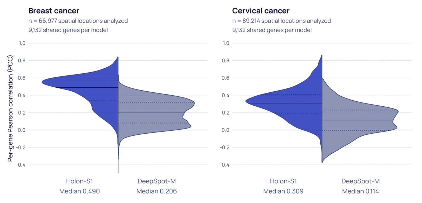 Holon-S1 beats DeepSpot-M on per-gene Pearson correlation for pathology-based gene expression prediction
Image: Sophont
Scanner robustness
Different slide scanners change the image, and a model that flips its answer with the scanner is hard to trust. Across Grundium, Hamamatsu and Leica scans of the same specimens, Holon-S1 showed lower normalized Wasserstein-1 distance than DeepSpot-M in 9 of 10 specimens (median 0.057 versus 0.120). The authors flag that this measures the consistency of the distribution, not the accuracy of the expression.
The report text refers to more than 15 scanner models and multiple organ-specific gene vocabularies; secondary summaries cite 16 organ types, which we could not confirm in the report. We verified the three-scanner experiment and the preparation comparison.
What did it find in real tumors?
The report includes several retrospective case studies. These are the part to read carefully, because they are hypotheses, not clinical results.
- Osteosarcoma. Using 11 initial diagnostic specimens from a public patient database, the model predicted higher expression of FAP, MDM2, KDR, CD276 and CLEC14A in tumor-like regions. Inferred MDM2 exceeded nine of 10 comparator patients. The model was not trained on osteosarcoma.
- Immune change over treatment. Post-treatment resections showed greater predicted immune infiltration than pre-treatment resections.
- Immune cell regions. On OrionCRC, classifying B-cell-dominant versus T-cell-dominant regions reached 88.5% held-out balanced accuracy and an AUROC of 0.943, checked against immunofluorescence ground truth.
- Triple-negative breast cancer. A spatial gradient of the gene EREG (called gEREG) predicted pathologic complete response in the held-out TransNEO cohort, and stayed significant after adjusting for stromal tumor-infiltrating lymphocytes, which did not predict response in that cohort.
- Colorectal cancer. Greater spatial segregation of proliferation and stroma/EMT programs was associated with better disease-specific survival, and the association held in the SurGen validation cohort.
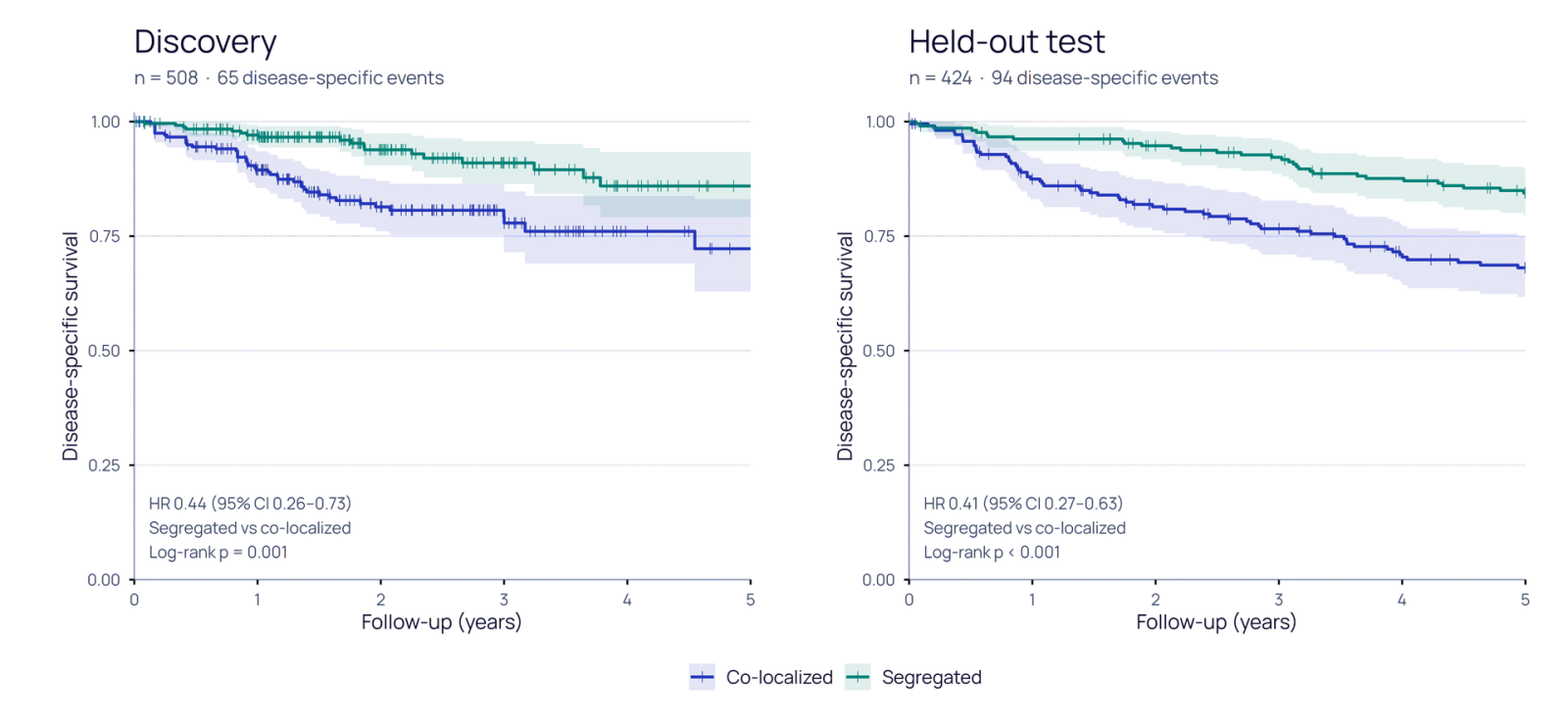 Holon-S1 predicted spatial programs stratify colorectal cancer disease-specific survival
Image: Sophont
Press summaries say the model "has already flagged potential new biomarkers" in colon and breast cancer. That is a fair description of the EREG and colorectal findings, as long as "potential" is read literally: they are retrospective associations in specific cohorts.
How to read the numbers without overreading them
Three habits help when you see a claim like "predicts gene expression from a slide."
First, ask what the correlation is computed over. The report uses median per-gene Pearson correlation between predicted and measured expression across held-out slides. A high median can coexist with many genes that are predicted poorly, which is why the authors also count how many genes pass increasing accuracy thresholds, grouped by assay technology and tissue type.
Second, ask which platform supplied the ground truth. The report notes that newer platforms such as Atera gave higher correlations than Xenium samples. That could mean the newer assay is less noisy, so the target itself is cleaner, rather than the model being better on those tissues. The authors acknowledge that tissue effects and platform effects cannot be fully separated in their held-out cohorts.
Third, separate prediction from clinical value. A gene-level correlation says the model recovers a pattern the assay also sees. A survival or treatment-response association says that pattern tracks an outcome in one cohort. Neither says a doctor should change treatment. The EREG result is the most clinically suggestive item in the report, and it is a held-out retrospective cohort in one breast cancer subtype.
Limitations the authors state
The report is candid about its limits, and they decide how far you should trust the headline.
- Regional, not single-cell. Predictions work at the regional level. Cell-level models are described as future work.
- Platform confounding. Held-out cohorts used different assay technologies, so tissue-specific effects cannot be fully separated from platform effects. That is why the authors use a confidence-based gene "vocabulary" for each organ.
- Retrospective evidence. Reproducibility and clinical utility need prospective and additional retrospective studies.
- Not a diagnostic. Research-only, not approved for patient diagnosis.
Where does it fit in the wider AI-for-biology wave?
Holon-S1 is one of several recent models that infer hidden biology from cheap data. We covered a genome language model that designed phage therapy for a patient, an AI model that reads a face photo to predict cancer survival, and Anthropic's protein binders against scorpion toxin, where only 17 of 211 designs held up in the lab. The common lesson is the same: results on benchmarks and cohorts are the start of validation, not the end.
Related imaging work includes Nvidia's Cosmos3-H Dreams surgical simulator and OpenAI's acquisition of Orchestra Bio, which show labs moving toward life-science tooling.
What this means for builders and researchers
If you build in digital pathology, three points are practical.
- Inputs are cheap. The pipeline starts from standard H&E whole-slide images, which many labs already have scanned.
- Benchmarks to check. The report names DeepSpot-M, Path2Space, HTAN, TransNEO, SurGen, OrionCRC and the PAR scanner dataset. If you evaluate a competing model, use the same sets, and note that Sophont chose them.
- Access is unclear. Sophont says it is seeking partnerships with research teams to integrate the technology into clinical workflows. The report page lists an "Explore Holon-S1" link and a PDF, and does not state licensing or weights availability in the text we read. Check Sophont directly before planning on running it.
Sophont previously published an open pathology foundation model recipe, OpenMidnight, so the lab has a record of releasing methods. Whether Holon-S1 weights will be released is not stated.
What is claimed and what is verified
| Claim | Status |
|---|---|
| Predicts spatial gene expression from H&E | Described in the technical report; authors' own evaluation |
| Beats DeepSpot-M and Path2Space | Reported by the authors on chosen datasets; no independent replication found |
| Many scanners, multiple organs, both tissue types | Report cites more than 15 scanner models; organ count not confirmed; we confirmed three scanners and the FFPE versus frozen test |
| Biomarker discovery in colon and breast cancer | Retrospective associations, not clinical validation |
| Ready for patients | No. Research-only |
Bottom line
Holon-S1 is an interesting, openly reasoned step toward cheap spatial molecular readouts, and its report is more cautious than the headlines about "avoiding costly tests." It does not replace molecular diagnostics today. The things to watch are independent replication, a prospective study of the EREG finding, single-cell resolution and a clear access policy. Follow @explainx_ai for updates.
Related reading
- Radical Numerics Omnii-1: a genome language model and an AI-designed phage
- Mass General Brigham FAHR: a face foundation model for cancer survival
- Claude-designed protein binders against scorpion toxin
- Nvidia Cosmos3-H Dreams surgical simulator
- OpenAI acquires Orchestra Bio
- Google Earth AI geospatial reasoning agent
- Official: Holon-S1 technical report, OpenMidnight from Sophont
Related: Hugging Face Carbon-X, an open chat model for DNA questions.
Details are accurate as of October 21, 2026; Holon-S1 is a research tool and nothing here is medical advice.